Ever wondered how your dentist sees what’s happening deep inside your teeth and gums? It’s not magic — it’s thanks to radiology in dentistry! In this blog, we’ll break down what radiology in dentistry is all about, how it has evolved, and why it’s such a valuable tool for maintaining your smile’s health and brightness. So, let’s dive in!
What is Radiology in Dentistry?
Simply put, radiology in dentistry is like having a special superpower that lets your dentist see things they can’t see with just their eyes. It involves using various imaging technologies, most commonly X-rays, to create images of your teeth, bones, and surrounding tissues. Think of it as a detailed map of your mouth, revealing hidden problems or confirming that everything is shipshape.
These pictures are incredibly important because many dental issues, such as cavities between teeth, infections in the bone, or even problems with the jaw, are often invisible on the surface. Without radiology, your dentist would be making guesses, and that’s not good for your dental health.
Types of Dental Radiology
Over the years, dental radiology has undergone significant evolution. Here are the main types you might encounter:
- Intraoral X-rays: These are the most common type, where the X-ray film or sensor is placed inside your mouth. You’ve probably had these before! They provide very detailed pictures of individual teeth and the surrounding bone.
- Bitewings: These show the crowns of your upper and lower back teeth in one image. They’re great for spotting cavities between teeth and checking the bone levels around your teeth.
- Periapical X-rays: These show an entire tooth, from its crown (the part you see) all the way to the tip of its root and the surrounding bone. They’re used to look for infections, problems with root canals, or issues with the bone supporting the tooth.
- Occlusal X-rays: These are taken with the film or sensor held between your teeth, showing a larger area of either the upper or lower jaw. They’re useful for seeing how teeth are developing, checking for extra teeth, or looking at larger areas of bone.
- Extraoral X-rays: These are taken with the X-ray machine outside your mouth. They give a broader view of your jaws and skull.
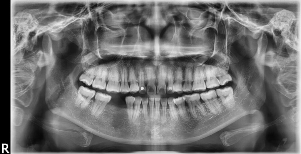
- Panoramic X-rays (OPG): This is a fantastic X-ray that captures a single image of your entire mouth – all your teeth, both jaws, and even parts of your sinuses and jaw joints (TMJ). It’s super useful for getting an overview of your oral health, checking for wisdom teeth, looking for cysts or tumours, or planning for orthodontic treatment. Many dentists, including a good Harrington Park dentist, will use an OPG as a standard part of a comprehensive dental check-up.
- Cephalometric X-rays: These are usually taken for orthodontic treatment. They show a side view of your head, allowing the dentist or orthodontist to measure the relationships between your teeth, jaws, and facial bones. This helps in planning how to straighten teeth and improve your bite.
- Cone Beam Computed Tomography (CBCT): This is a newer, very advanced type of X-ray that creates 3D images of your teeth, soft tissues, nerve pathways, and bone in a single scan. Think of it like a super-detailed 3D map of your mouth. CBCT is incredibly useful for complex cases, like planning dental implants, figuring out difficult wisdom tooth extractions, or diagnosing tricky infections. It provides so much more information than traditional 2D X-rays, allowing for much more precise treatment planning.
How Radiology Helps in Diagnosing Dental Conditions
Radiology is the detective in the world of dentistry. Without it, many dental problems would go unnoticed until they become painful or severe. Here’s how it helps your dentist diagnose various conditions:
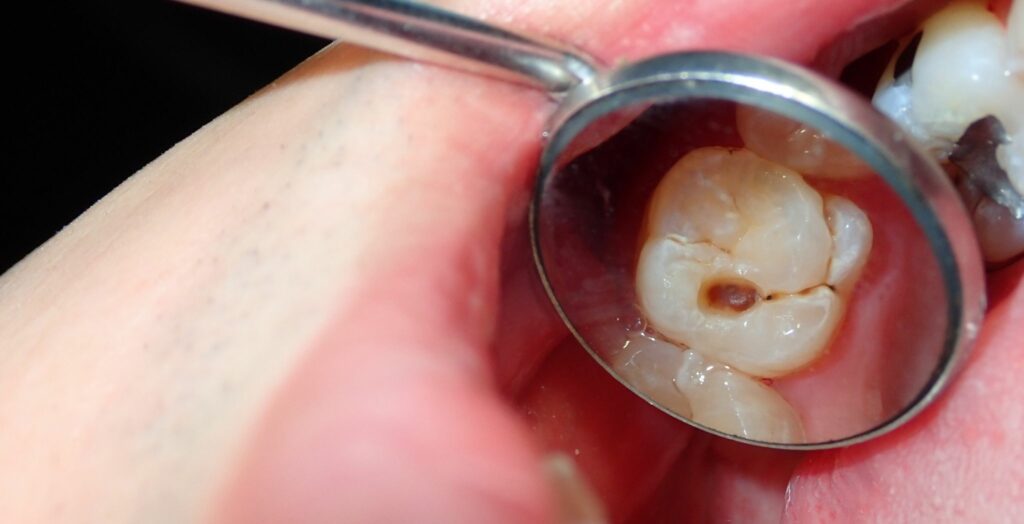
- Finding Cavities: While your dentist can see some cavities on the surface, many start in between your teeth or under old fillings, where they’re invisible. Bitewing X-rays are excellent at revealing these hidden cavities, allowing your dentist to treat them before they get bigger and cause more damage.
- Spotting Infections: Infections can develop at the tip of a tooth’s root or in the surrounding bone, often without any pain in the early stages. Periapical X-rays can show these infections as dark areas around the root, indicating inflammation or bone loss. Catching these early can save your tooth!
- Checking Bone Health: The bone that supports your teeth is crucial. Periodontal disease (gum disease) can cause this bone to wear away. X-rays can show the level of bone support around your teeth, helping your dentist assess the severity of gum disease and plan appropriate treatment.
- Evaluating Wisdom Teeth: Wisdom teeth can cause a lot of trouble, from becoming impacted (stuck) to causing pain or infection. Panoramic X-rays are essential for seeing the position of your wisdom teeth, if they’re impacted, and if they’re likely to cause problems in the future. This helps your dentist decide if they need to be removed.
- Detecting Cysts and Tumours: While rare, cysts and tumours can develop in the jawbones or soft tissues of the mouth. X-rays, especially panoramic and CBCT scans, can help identify these abnormalities, allowing for early diagnosis and treatment.
- Assessing Jaw Problems: If you have pain in your jaw joint (TMJ) or problems with jaw movement, X-rays can provide valuable information about the joint’s structure and any underlying issues.
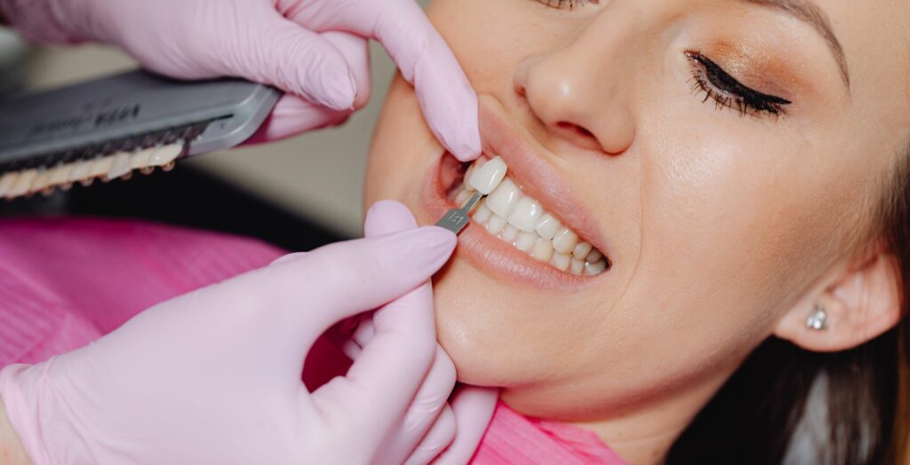
- Planning Orthodontic Treatment: For those considering braces or other orthodontic treatments, X-rays like panoramic and cephalometric views are vital. They help the orthodontist understand the relationship between your teeth and jaws, allowing them to create a precise treatment plan to straighten your smile.
The Role of X-rays in Preventing Dental Issues
It’s not just about finding problems; X-rays play a huge role in preventing them too! By allowing your dentist to see what’s happening beneath the surface, they can often catch issues in their very early stages, before they become big, painful, and expensive problems.
For example, a tiny cavity between your teeth might not cause any pain, but an X-ray can spot it. Treating that small cavity is much easier, quicker, and less invasive than waiting for it to grow larger, cause a toothache, and potentially require a root canal or extraction. Similarly, detecting early bone loss due to gum disease on an X-ray allows your dentist to intervene and halt the disease’s progression, thereby saving your teeth from becoming loose or falling out.
Regular X-rays, as recommended by your dentist, are a key part of preventative dentistry. They help your dentist stay ahead of potential issues, ensuring your oral health remains in top condition.
Benefits of Digital Radiology
For many years, dental X-rays used film, similar to old cameras. However, just as photography has transitioned to digital, so has dental radiology. Digital radiology is now the standard in most modern dental practices, and it comes with a heap of fantastic benefits:
- Reduced Radiation Exposure: This is a big one! Digital X-ray sensors are significantly more sensitive than traditional film, requiring substantially less radiation to produce a clear image. This significantly reduces your exposure to radiation, making the procedure even safer. Your dentist, like dentist Leppington will prioritise your safety, and digital X-rays are a key part of that.
- Instant Images: No more waiting for films to develop! Digital X-rays appear almost instantly on a computer screen. This means your dentist can show you the images right away and explain what they see, making the consultation more interactive and understandable.
- Improved Image Quality: Digital images can be enhanced, zoomed in, and adjusted for brightness and contrast. This allows your dentist to see details that might be harder to spot on traditional film, leading to more accurate diagnoses.
- Easier Storage and Sharing: Digital images are stored electronically, making them easy to access, share with specialists (if needed), and keep track of your dental history. No more lost films!
- Environmentally Friendly: Digital radiology eliminates the need for harmful chemicals used in film development, making it a much greener option.
Safety Considerations with Dental Radiology
It’s natural to have questions about safety when it comes to X-rays. It’s essential to remember that the amount of radiation from dental X-rays is extremely small, especially with modern digital systems. You actually receive more radiation exposure from everyday activities, such as flying in an aeroplane or simply being outdoors in the sun.
Your dentist is highly trained to use X-ray equipment safely and will only take X-rays when necessary. They follow strict guidelines to minimise your exposure, including:
- Using Lead Aprons and Thyroid Collars: These protective covers shield your body from any scattered radiation.
- Using High-Speed Film or Digital Sensors: As mentioned, digital systems significantly reduce radiation dose.
- Proper Positioning: The X-ray machine is carefully positioned to target only the area of interest, limiting exposure to other parts of your body.
- Following the “As Low As Reasonably Achievable” (ALARA) Principle: This means dentists only take the X-rays that are absolutely needed for diagnosis and treatment planning, using the lowest possible dose.
If you’re pregnant or think you might be, it’s very important to tell your dentist. While dental X-rays are generally considered safe during pregnancy with proper precautions, your dentist may choose to postpone non-urgent X-rays.
What to Expect During a Dental Radiology Procedure
A dental radiology procedure is quick and painless! Here’s a general idea of what to expect:
- Preparation: You’ll be asked to remove any glasses, jewellery, or removable dental appliances that might interfere with the X-ray image. You’ll then be given a lead apron and possibly a thyroid collar to wear for protection.
- Positioning: Depending on the type of X-ray, you’ll either sit in the dental chair or stand in front of a special machine. For intraoral X-rays, a small film or digital sensor will be placed inside your mouth, and you’ll be asked to bite down gently. For panoramic X-rays, you’ll stand still while a rotating arm moves around your head. For CBCT scans, you might sit or stand while the scanner rotates around you.
- Taking the Image: The dental assistant or dentist will step out of the room or behind a protective barrier while the X-ray is taken. You’ll be asked to stay very still for a few seconds. You won’t feel anything during the process.
- Review: For digital X-rays, the images will appear on the computer screen almost immediately. Your dentist will then review them with you, explaining their findings and discussing any necessary treatment.
The entire process typically takes only a few minutes, depending on the number and type of X-rays required.
Radiology and Treatment Planning
Beyond diagnosis, radiology plays a crucial role in effective treatment planning. Once your dentist has a clear picture of your oral health from the X-rays, they can create the best possible plan for your care.
- Fillings and Crowns: X-rays show the extent of cavities, helping your dentist decide if a filling is enough or if a crown is needed.
- Root Canals: If a tooth needs a root canal, X-rays guide the dentist in understanding the root anatomy and ensuring the entire infection is cleared.
- Dental Implants: For dental implants, CBCT scans are invaluable. They show the exact amount and quality of bone available, as well as the location of nerves and sinuses, allowing for precise implant placement and reducing the risk of complications.
- Extractions: Especially for wisdom teeth or complex extractions, X-rays help your dentist understand the position of the tooth, its roots, and its relationship to nearby nerves, making the extraction safer and more predictable.
- Orthodontics: As mentioned, X-rays provide the detailed measurements needed to plan the movement of teeth and jaws for braces or aligners.
- Oral Surgery: Any oral surgery procedure relies heavily on detailed X-ray imaging to ensure safety and precision.
Conclusion
Radiology in dentistry is a true game-changer. It’s transformed the way dentists diagnose, prevent, and treat dental problems, all for the better. From finding hidden cavities to planning complex surgeries, the information provided by dental X-rays is invaluable. With the advancements in digital technology, dental radiology is safer, faster, and more effective than ever before.
So, the next time your dentist suggests an X-ray, you’ll know exactly why it’s such an important part of keeping your smile healthy and bright. It’s all about providing you with the best possible care, ensuring that hidden issues are spotted early and treated effectively.
Ready to Keep Your Smile in Top Shape?
Whether you’re in Leppington or Harrington Park, finding a dentist who embraces advanced dental radiology means you’re getting the most thorough and modern care available.
Don’t put off your next dental check-up – healthy teeth and gums start with a clear picture!